
Page 1 - Renal Colic
P. 1
Urological Health
Renal colic
A kidney stone may cause severe pain requiring urgent medical attention.
ou have been found to have a kidney stone The pain occurs on the side of the stone but its
Ycausing pain (renal colic). A kidney is found precise location depends upon where the stone
in each flank under the ribs. The kidneys play an becomes lodged in the kidney or ureter. The nature
important role in eliminating waste products from the and location of the pain may change as the stone
body. These waste products normally remain dissolved migrates down the ureter toward the bladder. Renal
in the urine as it passes from the kidney through its colic will often start in the flank (between the ribs and
drainage system (calyces, renal pelvis and ureter) into hip) or lower back but it can also be felt in the lower
the bladder. abdomen, groin, genitals or inner thigh. The pain of
renal colic may be associated with nausea, vomiting,
Kidney stones are crystalline particles that form in the and frequent or urgent urges to urinate, which may
urine, often producing pain when they obstruct urine be painful. Blood in the urine (hematuria) occurs
drainage from a kidney. About one in ten Canadians frequently with kidney stones. This blood may be
will develop a kidney stone, and of these, half will form visible or microscopic.
more than one over their lifetime. This problem is more
common in men than in women, and, it occurs rarely The diagnosis of renal colic may be suspected by
in children. the description of pain experienced supported
by blood and urine tests. Some type of medical
imaging is necessary to confirm the diagnosis and
determine the size and location of the kidney stone.
A CT scan is the most commonly used imaging test to
diagnose a kidney stone and determine its size and
location. Other tests may include ultrasound imaging
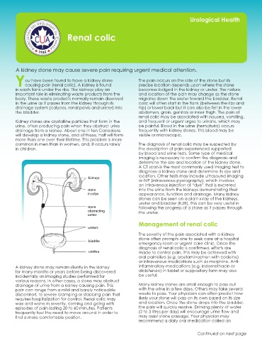
kidneys
or IVP (intravenous pyelography), which involves
an intravenous injection of “dye” that is excreted
stone into the urine from the kidneys demonstrating their
in calyx appearance, function and drainage. Many kidney
stones can be seen on a plain x-ray of the kidneys,
ureter and bladder (KUB). This can be very useful in
stone following the progress of a stone as it passes through
obstructing the ureter.
ureter
Management of renal colic
ureters
The severity of the pain associated with a kidney
stone often prompts one to seek care at a hospital
bladder
emergency room or urgent care clinic. Once the
diagnosis of renal colic is confirmed, efforts are
urethra made to control pain. This may be achieved with
oral painkillers (e.g. acetaminophen with codeine)
or intravenous medications such as morphine. Anti-
A kidney stone may remain silently in the kidney inflammatory medications (e.g. indomethacin or
for many months or years before being discovered diclofenac) in tablet or suppository form may also
incidentally on imaging studies performed for be useful.
various reasons. In other cases, a stone may obstruct
drainage of urine from a kidney causing pain. This Many kidney stones are small enough to pass out
pain can range from a mild and barely noticeable with the urine in a few days. Others may take several
discomfort, to severe cramping or stabbing pain that weeks to pass. Your physician can often predict how
requires hospitalization for control. Renal colic may likely your stone will pass on its own based on its size
wax and wane in severity, coming and going with and location. Once the stone drops into the bladder,
episodes of pain lasting 20 to 60 minutes. Patients the pain will quickly resolve. Drinking plenty of water
frequently feel the need to move around in order to (2 to 3 litres per day) will encourage urine flow and
find a more comfortable position. may assist stone passage. Your physician may
recommend a daily oral medication called an
Continued on next page