
Page 14 - Diagnosis, management, and surveillance of neurogenic lower urinary tract dysfunction – Full text
P. 14
Kavanagh et al
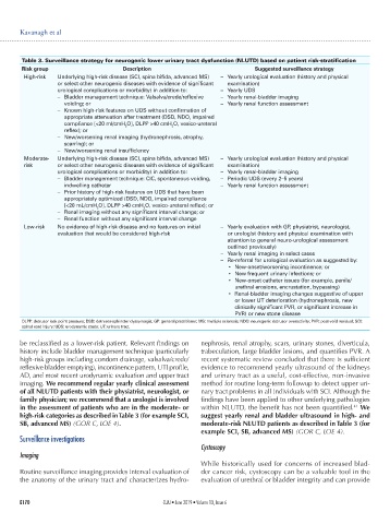
Table 3. Surveillance strategy for neurogenic lower urinary tract dysfunction (NLUTD) based on patient risk-stratification
Risk group Description Suggested surveillance strategy
High-risk Underlying high-risk disease (SCI, spina bifida, advanced MS) – Yearly urological evaluation (history and physical
or select other neurogenic diseases with evidence of significant examination)
urological complications or morbidity) in addition to: – Yearly UDS
– Bladder management technique: Valsalva/crede/reflexive – Yearly renal-bladder imaging
voiding; or – Yearly renal function assessment
– Known high-risk features on UDS without confirmation of
appropriate attenuation after treatment (DSD, NDO, impaired
compliance [<20 ml/cmH O], DLPP >40 cmH O, vesico-ureteral
2
2
reflex); or
– New/worsening renal imaging (hydronephrosis, atrophy,
scarring); or
– New/worsening renal insufficiency
Moderate- Underlying high-risk disease (SCI, spina bifida, advanced MS) – Yearly urological evaluation (history and physical
risk or select other neurogenic diseases with evidence of significant examination)
urological complications or morbidity) in addition to: – Yearly renal-bladder imaging
– Bladder management technique: CIC, spontaneous voiding, – Periodic UDS (every 2–5 years)
indwelling catheter – Yearly renal function assessment
– Prior history of high-risk features on UDS that have been
appropriately optimized (DSD, NDO, impaired compliance
[<20 mL/cmH O], DLPP >40 cmH O, vesico-ureteral reflex); or
2
2
– Renal imaging without any significant interval change; or
– Renal function without any significant interval change
Low-risk No evidence of high-risk disease and no features on initial – Yearly evaluation with GP, physiatrist, neurologist,
evaluation that would be considered high-risk or urologist (history and physical examination with
attention to general neuro-urological assessment
outlined previously)
– Yearly renal imaging in select cases
– Re-referral for urological evaluation as suggested by:
• New-onset/worsening incontinence; or
• New frequent urinary infections; or
• New-onset catheter issues (for example, penile/
urethral erosions, encrustation, bypassing)
• Renal-bladder imaging changes suggestive of upper
or lower UT deterioration (hydronephrosis, new
clinically significant PVR, or significant increase in
PVR) or new stone disease
DLPP: detrusor leak point pressure; DSD: detrusor-sphincter dyssynergia; GP: general practitioner; MS: multiple sclerosis; NDO: neurogenic detrusor overactivity; PVR: post-void residual; SCI:
spinal cord injury; UDS: urodynamic study; UT: urinary tract.
be reclassified as a lower-risk patient. Relevant findings on nephrosis, renal atrophy, scars, urinary stones, diverticula,
history include bladder management technique (particularly trabeculation, large bladder lesions, and quantifies PVR. A
high-risk groups including condom drainage, valsalva/crede/ recent systematic review concluded that there is sufficient
reflexive bladder emptying), incontinence pattern, UTI profile, evidence to recommend yearly ultrasound of the kidneys
AD, and most recent urodynamic evaluation and upper tract and urinary tract as a useful, cost-effective, non-invasive
imaging. We recommend regular yearly clinical assessment method for routine long-term followup to detect upper uri-
of all NLUTD patients with their physiatrist, neurologist, or nary tract problems in all individuals with SCI. Although the
family physician; we recommend that a urologist is involved findings have been applied to other underlying pathologies
41
in the assessment of patients who are in the moderate- or within NLUTD, the benefit has not been quantified. We
high-risk categories as described in Table 3 (for example SCI, suggest yearly renal and bladder ultrasound in high- and
SB, advanced MS) (GOR C, LOE 4). moderate-risk NLUTD patients as described in Table 3 (for
example SCI, SB, advanced MS) (GOR C, LOE 4).
Surveillance investigations
Cystoscopy
Imaging
While historically used for concerns of increased blad-
Routine surveillance imaging provides interval evaluation of der cancer risk, cystoscopy can be a valuable tool in the
the anatomy of the urinary tract and characterizes hydro- evaluation of urethral or bladder integrity and can provide
E170 CUAJ • June 2019 • Volume 13, Issue 6