
Page 1 - Ureteropelvic Junction Obstruction
P. 1
Urological Health
Ureteropelvic junction
obstruction
A ureteropelvic junction obstruction is a partial blockage of the drainage system of the kidney.
our kidneys filter blood to eliminate waste
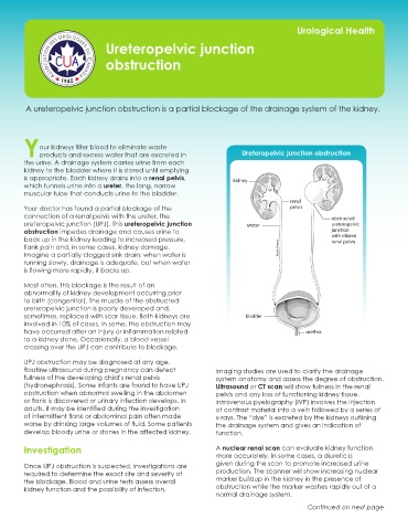
Yproducts and excess water that are excreted in Ureteropelvic junction obstruction
the urine. A drainage system carries urine from each
kidney to the bladder where it is stored until emptying
is appropriate. Each kidney drains into a renal pelvis, kidney
which funnels urine into a ureter, the long, narrow
muscular tube that conducts urine to the bladder.
renal
Your doctor has found a partial blockage of the pelvis
connection of a renal pelvis with the ureter, the obstructed
ureteropelvic junction (UPJ). This ureteropelvic junction ureter ureteropelvic
obstruction impedes drainage and causes urine to junction
back up in the kidney leading to increased pressure, with dilated
renal pelvis
flank pain and, in some cases, kidney damage. Roula Drossis
Imagine a partially clogged sink drain: when water is
running slowly, drainage is adequate, but when water
is flowing more rapidly, it backs up.
Most often, this blockage is the result of an
abnormality of kidney development occurring prior
to birth (congenital). The muscle of the obstructed
ureteropelvic junction is poorly developed and,
sometimes, replaced with scar tissue. Both kidneys are bladder
involved in 10% of cases. In some, the obstruction may
have occurred after an injury or inflammation related urethra
to a kidney stone. Occasionally, a blood vessel
crossing over the UPJ can contribute to blockage. Ureteropelvic Junction Obstruction
UPJ obstruction may be diagnosed at any age.
Routine ultrasound during pregnancy can detect Imaging studies are used to clarify the drainage
fullness of the developing child’s renal pelvis system anatomy and assess the degree of obstruction.
(hydronephrosis). Some infants are found to have UPJ Ultrasound or CT scan will show fullness in the renal
obstruction when abnormal swelling in the abdomen pelvis and any loss of functioning kidney tissue.
or flank is discovered or urinary infection develops. In Intravenous pyelography (IVP) involves the injection
adults, it may be identified during the investigation of contrast material into a vein followed by a series of
of intermittent flank or abdominal pain often made x-rays. The “dye” is excreted by the kidneys outlining
worse by drinking large volumes of fluid. Some patients the drainage system and gives an indication of
develop bloody urine or stones in the affected kidney. function.
Investigation A nuclear renal scan can evaluate kidney function
more accurately. In some cases, a diuretic is
Once UPJ obstruction is suspected, investigations are given during the scan to promote increased urine
required to determine the exact site and severity of production. The scanner will show increasing nuclear
the blockage. Blood and urine tests assess overall marker buildup in the kidney in the presence of
kidney function and the possibility of infection. obstruction while the marker washes rapidly out of a
normal drainage system.
Continued on next page