
Page 2 - Ureteropelvic Junction Obstruction
P. 2
Ureteropelvic junction obstruction
Your urologist may recommend a cystoscopy and
retrograde pyelography. This involves passing a
narrow visualizing instrument (scope) through the
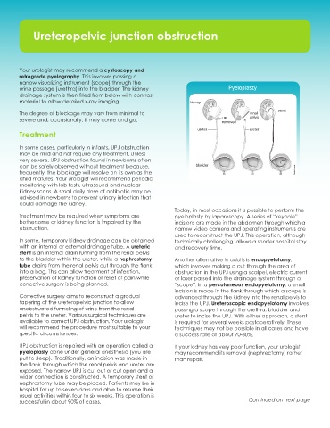
urine passage (urethra) into the bladder. The kidney Pyeloplasty
drainage system is then filled from below with contrast
material to allow detailed x-ray imaging. kidney
The degree of blockage may vary from minimal to renal stent
severe and, occasionally, it may come and go. UPJ pelvis
removed
ureter ureter
Treatment
In some cases, particularly in infants, UPJ obstruction
may be mild and not require any treatment. Unless
very severe, UPJ obstruction found in newborns often
can be safely observed without treatment because, bladder
frequently, the blockage will resolve on its own as the Roula Drossis
child matures. Your urologist will recommend periodic Pyeloplasty
monitoring with lab tests, ultrasound and nuclear
kidney scans. A small daily dose of antibiotic may be
advised in newborns to prevent urinary infection that
could damage the kidney.
Today, in most occasions it is possible to perform the
Treatment may be required when symptoms are pyeloplasty by laparoscopy. A series of “keyhole”
bothersome or kidney function is impaired by the incisions are made in the abdomen through which a
obstruction. narrow video camera and operating instruments are
used to reconstruct the UPJ. This operation, although
In some, temporary kidney drainage can be obtained technically challenging, allows a shorter hospital stay
with an internal or external drainage tube. A ureteric and recovery time.
stent is an internal drain running from the renal pelvis
to the bladder within the ureter, while a nephrostomy Another alternative in adults is endopyelotomy,
tube drains from the renal pelvis out through the flank which involves making a cut through the area of
into a bag. This can allow treatment of infection, obstruction in the UPJ using a scalpel, electric current
preservation of kidney function or relief of pain while or laser passed into the drainage system through a
corrective surgery is being planned. “scope”. In a percutaneous endopyelotomy, a small
incision is made in the flank through which a scope is
Corrective surgery aims to reconstruct a gradual advanced through the kidney into the renal pelvis to
tapering of the ureteropelvic junction to allow incise the UPJ. Ureteroscopic endopyelotomy involves
unobstructed funneling of urine from the renal passing a scope through the urethra, bladder and
pelvis to the ureter. Various surgical techniques are ureter to incise the UPJ. With either approach, a stent
available to correct UPJ obstruction. Your urologist is required for several weeks postoperatively. These
will recommend the procedure most suitable to your techniques may not be possible in all cases and have
specific circumstances. a success rate of about 70-80%.
UPJ obstruction is repaired with an operation called a If your kidney has very poor function, your urologist
pyeloplasty done under general anesthesia (you are may recommend its removal (nephrectomy) rather
put to sleep). Traditionally, an incision was made in than repair.
the flank through which the renal pelvis and ureter are
exposed. The narrow UPJ is cut out or cut open and a
wider connection is constructed. A temporary stent or
nephrostomy tube may be placed. Patients may be in
hospital for up to seven days and able to resume their
usual activities within four to six weeks. This operation is
successful in about 90% of cases. Continued on next page