
Page 20 - Urological Health
P. 20
Tests: Your doctor may suspect renal colic because of the pain you describe and by simple blood and
urine tests, and by imaging tests to get a detailed picture of your kidney, ureter and bladder (simple X-
ray called a kidney/ureter/bladder [KUB] or computed tomography [CT] scan) to determine the size
and location of the kidney stone. A stone-protocol CT scan, which uses less radiation than a normal
CT scan, is common, although an ultrasound may also be used. Another rare test is the dye test
(intravenous pyelogram [IVP]), which is another type of X-ray that takes pictures of the urinary tract
after a dye is inserted. Many kidney stones are seen on a KUB X-ray – this is a useful test that allows
your doctor to follow the progress of the stone through the ureter.
Treatment: The severity of your pain due to the kidney stone will often bring you to the ER. Once the
doctor confirms the diagnosis, your pain can be controlled with oral painkillers (like acetaminophen
with codeine) or intravenous medications, such as morphine. Anti-inflammatory medications (like
indomethacin or diclofenac) in tablet or suppository form (in the rectum) may also be useful. Many
kidney stones are small enough to pass out of your body on their own with the urine in a few days.
You can also wait for the stone to pass (also called expectant therapy). If you are taking this route, you
will need pain relief, an antispasmodic agent and/or anti-inflammatory drugs, adequate hydration
and antibiotics (if there are signs of a urinary infection). Drink plenty of water (2 to 3 litres per day);
this will make you go to the bathroom and may help pass the stone. Your doctor may recommend a
daily oral medication called an alpha-blocker (e.g., tamsolusin or Flomax) to relax your ureter muscles
to make the stone passage easier.
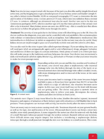
Depending on how sick you are and the size, number and location of
the stones, your doctor may place a nephrostomy tube (external
drainage tube into the kidney through the skin of the back) or an
internal ureteral stent (internal drainage tube called a double J stent),
with stone disintegration and/or removal of the stone, at the same
time or later.
If your pain becomes hard to manage, if the stone becomes lodged
and fails to pass, or if you have fever (greater than 38.5ºC) or have the
chills (which is a sign of infection), the situation becomes more
urgent. In this case, your stone itself may not be dealt with because
you are getting sicker. The doctor may place a ureteric stent or
nephrostomy tube to relieve your pain, decompress the urinary
Figure 2. A ureteric stent. system and allow the kidney to drain urine.
The ureteric stent (Figure 2) may cause blood in the urine, bladder discomfort (spasm), increased
frequency and urgency of urination or flank (kidney) pain with urination or a full bladder due to back
pressure. These symptoms can increase with activity, but resolve shortly after the stent is removed.
There are many ways to treat a stone causing renal colic. Treatment depends on your general health,
as well as the type, size and location of the stone. Ultrasound shock waves can break a stone into
smaller, more easily passed pieces. A stone trapped in the ureter can often be removed with the help
of a small fiberoptic telescope passed through the urethra (urinary channel) without any incisions.
More difficult stones may require surgery that includes a introducing a nephroscope (kidney
telescope) through a small puncture through the skin over the kidney or a small incision. This is rare.
19