
Page 83 - CUA Absracts 2022_Fulldraft
P. 83
Poster 8: Endourology, Renal Transplant
incidental finding of hepatic steatosis, steatosis progression was noted the TFL group. Operative time was calculated from scope introduction
for 13 (17.1%) and regression was noted for two (2.6%). Twelve patients to removal. SFR was assessed with one or multiple imaging modalities;
were referred to hepatology for further liver investigations. Among these non-contrast computed tomography (CT), kidney-bladder-ureter (KUB)
patients, one (8.3%) had cirrhosis, two (16.7%) had fibrosis, and two ultrasound, or X-ray. Other variables, including stone size, density, and
(16.7%) had moderate-to-severe steatosis. prior ureteral stenting, were recorded. In the case of multiple stones, the
Conclusions: The incidental finding of fatty liver on ultrasound of patients total stone surface area was measured.
followed for nephrolithiasis is common, especially in overweight or Results: Patient age (years), prior ureteral stenting, procedure time
smoker patients. While only a small proportion of these patients have (min:sec), and total stone surface area were similar between groups.
significant fibroscan-confirmed fibrosis or cirrhosis, urologists could initi- The number of treated stones was higher in the TFL group (p=0.0015).
ate lifestyle changes that improve outcomes for both liver and kidney Compared to Ho:YAG, TFL showed a significantly higher rate of stone
stone diseases and refer to gastroenterology/hepatology when appropriate. fragmentation per mm stone surface area (p=0.02). The data showed
2
a similar SFR between Ho:YAG and TFL groups, as well as the size of
MP-8.9 residual fragments (<4 mm or >4mm) (Table 1).
Conclusions: This study demonstrates that the TFL has a more efficient
Thulium fiber laser vs. holmium:YAG: A clinical comparison of lithotripsy effect per mm stone surface area than Ho:YAG. SFR was similar
2
laser lithotripsy efficiency in a retrospective of 73 patients at a for both Ho:YAG and TFL; however, this is confounded by the fact that a
tertiary stone center significantly higher number of stones have been treated with TFL. In our
Alec Mitchell , Victor Wong , Abdulghafour Halawani , Ryan Paterson , study, TFL produced a similar SFR to Ho:YAG at a more efficient rate of
1
1
1
1
Ben Chew 1 lithotripsy. Further clinical studies are warranted to tease out the above
1 Department of Urologic Sciences, University of British Columbia, results and to determine whether thulium can truly challenge holmium
Vancouver, BC, Canada as the default laser in urology.
Introduction: Since its first use by Dr. Denstedt in 1993, the holmium
(Ho):YAG laser has been the gold standard laser for lithotripsy. The thul-
ium fiber laser (TFL) is a new laser technology that has shown promising MP-8.10
results in several preclinical studies. This new technology may expand the Retrograde ureteral stent vs. percutaneous nephrostomy tube
boundaries of laser lithotripsy. This study aimed to compare the efficacy drainage for obstructive urolithiasis: Predictors for spontaneous
of TFL and Ho:YAG in terms of stone fragmentation rate, operative time, stone passage
and stone-free rate (SFR). Abdulghafour Halawani 1,2,3 , Mudhar Hasan 4,5
Methods: A retrospective analysis was conducted at a tertiary stone center 1 Department of Urologic Sciences, The University of British Columbia,
to identify patients treated with Ho:YAG or TFL laser lithotripsy. Seventy- Vancouver, BC, Canada; Department of Urology, King Abdulaziz
2
three cases were included: 42 patients in the Ho:YAG group and 31 in University, Jeddah, Saudi Arabia; Department of Urology, Sodersjukhuset,
3
Karolinska Institutet, Stockholm, Sweden; Department of Urology,
4
Danderyds University Hospital, Stockholm, Sweden; Mediclinic City
5
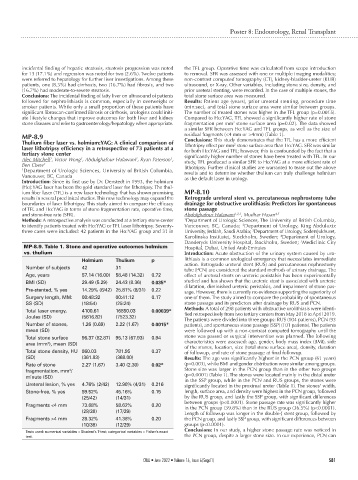
MP-8.9. Table 1. Stone and operative outcomes holmium Hospital, Dubai, United Arab Emirates
vs. thulium Introduction: Acute obstruction of the urinary system caused by uro-
Holmium Thulium p lithiasis is a common urological emergency that necessitates immediate
Number of subjects 42 31 action. Retrograde ureteral stent (RUS) and percutaneous nephrostomy
tube (PCN) are considered the standard methods of urinary drainage. The
Age, years 57.14 (16.00) 58.48 (14.32) 0.72 effect of ureteral stents on ureteric peristalsis has been experimentally
BMI (SD) 29.49 (5.29) 24.43 (8.36) 0.035* studied and has shown that the ureteric stent is associated with ureteric
dilatation, diminished ureteric peristalsis, and impairment of stone pas-
Pre-stented, % yes 14.29% (6/42) 25.81% (8/31) 0.22 sage. However, there is currently no evidence supporting the superiority of
Surgery length, MM: 00:48:32 00:41:12 0.17 one of them. The study aimed to compare the probability of spontaneous
SS (SD) (18:54) (25:24) stone passage and its predictors after drainage by RUS and PCN.
Total laser energy, 4100.61 16850.03 0.00039* Methods: A total of 298 patients with obstructive urolithiasis were identi-
fied retrospectively from two tertiary centers from May 2018 to April 2019.
Joules (SD) (5616.82) (7523.32) The patients were divided into three groups: RUS (104 patients), PCN (93
Number of stones, 1.26 (0.69) 2.22 (1.67) 0.0015* patients), and spontaneous stone passage (SSP) (101 patients). The patients
mean (SD) were followed up with a non-contrast computed tomography until the
Total stone surface 96.37 (82.87) 95.13 (67.93) 0.94 stone was passed or surgical intervention was planned. The following
area (mm ), mean (SD) characteristics were assessed: age, gender, body mass index (BMI), side
2
of the stones, location, size (total stone surface area), density, duration
Total stone density, HU 860.03 781.95 0.37 of followup, and rate of stone passage at final followup.
(SD) (361.83) (368.00) Results: The age was significantly highest in the PCN group (61 years)
Rate of stone 2.27 (1.67) 3.40 (2.30) 0.02* (p=0.003), while BMI and gender distribution were similar among groups.
fragmentation, mm / Stone size was larger in the PCN group than in the other two groups
2
minute (SD) (p=0.0001) (Table 1). The stones were located mainly in the distal ureter
in the SSP group, while in the PCN and RUS groups, the stones were
Ureteral lesion, % yes 4.76% (2/42) 12.90% (4/31) 0.216 significantly located in the proximal ureter (Table 1). The stones’ width,
Stone-free, % yes 59.52% 45.16% 0.15 length, surface area, and density were highest in the PCN group, followed
(25/42) (14/31) by the RUS group, and lastly the SSP group, with significant differences
Fragments <4 mm 73.68% 58.62% 0.20 between groups (p<0.0001). Stone passage rate was significantly higher
in the PCN group (39.8%) than in the RUS group (36.5%) (p<0.0001).
(28/38) (17/29) Length of followup was longer in the double-J stent group, followed by
Fragments >4 mm 28.32% 41.38% 0.20 the PCN group, and lastly SSP group, with significant differences between
(10/38) (12/29) groups (p<0.0001).
Tests used: numerical variables = Student’s T-test; categorical variables = Fisher’s exact Conclusions: In our study, a higher stone passage rate was noticed in
test. the PCN group, despite a larger stone size. In our experience, PCN can
CUAJ • June 2022 • Volume 16, Issue 6(Suppl1) S81