
Page 2 - CUA Best Practice Report: Diagnosis and management of radiation-induced hemorrhagic cystitis
P. 2
Goucher et al
rare, it is hypothesized that indirect damage occurs through terminology has led to the development of the Common
4
the creation of free radicals. Histological studies have dem- Terminology Criteria for Adverse Events as a uniform lexicon
onstrated increased proliferation of the urothelium in the for description of cancer-related adverse effects. 12
months following radiation. Damage to tight cellular junc-
tions and the loss of the normal polysaccharide layer allow Initial management
for increased permeability of urine bacteria and metabolites
1
causing increased damage to the underlying tissue. This
altered permeability of the urothelial cell layer has been Diagnosis and early assessment
demonstrated to be involved in late-stage radiation changes
5
in rat models and is hypothesized to play a large role in the In patients presenting with hematuria post-radiation, a thor-
development of post-radiation urinary symptoms. 6 ough assessment is needed to rule out secondary causes
Diffuse mucosal edema is noted in biopsies taken imme- before a diagnosis of RHC can be made. In a study explor-
diately post-radiation. This is followed by development of ing cystoscopic evaluation of 185 men treated with brachy-
vascular telangiectasia, submucosal hemorrhage, and inter- therapy for prostate cancer who presented with either macro-
stitial fibrosis. Subendothelial proliferation, edema, and scopic hematuria, microscopic hematuria, or persistent lower
medial thickening may progressively deplete the blood sup- urinary tract, 9.6% were found to have a new bladder tumour
ply to urothelium, resulting in endarteritis obliterans causing compared to 7% who were found to have radiation cystitis. 13
acute and chronic ischemia. These ischemic and necrotic While the majority of these symptomatic post-brachythera-
3
changes are proposed to give rise to subsequent develop- py patients had cystoscopies reported as normal (63.8%), a
ment of revascularization with superficial, fragile vessels that clinically significant number did have an observable etiology.
are responsible for bleeding in radiation cystitis. 4 Assessment should begin with a detailed history character-
Hospitalizations for RHC can be lengthy and costly. A izing the symptoms and confirming the history and treatment
recent retrospective study assessing 1111 patients admitted plan of a patient’s radiation therapy. Physical exams, includ-
for RHC in 2013 showed the median cost associated with ing an abdominal and pelvic exam to assess for alternative
each admission to be $7157 USD. This number rose to causes of bleeding, should be included. Laboratory tests,
$11 100 for those with hematuria severe enough to merit including a complete blood count, coagulation studies, serum
endoscopic evaluation/treatment. Multiple studies have creatinine, urinalyses, urine culture and cytology, should be
7
demonstrated the significant effect its protracted and recur- initiated. As with any patient presenting with hematuria and
rent nature can have on patient-rated quality of life scales. 8 a high risk of malignancy, all patients should undergo axial
There exists little consensus of how to best treat RHC, and imaging, preferably a computed tomography (CT)-urogram
previous surveys of practicing urologist have shown a lack to assess for upper tract sources of bleeding, and should also
of awareness of treatment options available. 9 undergo cystoscopic evaluation and biopsies of lesions con-
cerning for malignancy. Mild symptoms may resolve with
Classifications continuous bladder irrigation with saline solution and this
should be tried first in all patients with hematuria associated
The European Organization for Research and Treatment of with clotting or retention.
Cancer/Radiation Therapy Oncology Group (EORTC/RTOG) Recommendation: Assessment of a patient complain-
classification of late radiation effects is a commonly used clas- ing of hematuria post-radiation therapy should identify
sification system for grading of RHC (Table 1). It describes or exclude other pathological factors that may explain or
10
a combination of clinical and cystoscopic criteria for report- contribute to the patient’s symptoms (Grade 4C).
ing late radiation effects. The Late Effects of Normal Tissues
(LEBT)/SOMA scale has also been developed and uses a more Cystoscopic evaluation
complex combination of subjective, objective, management,
11
and analytical factors into radiation effect classification. A Cystoscopy in patients with new-onset or suspected RHC
more recent move to replace these systems with a common can be both diagnostic and therapeutic. The appearance of
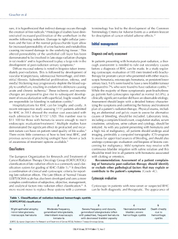
Table 1. Classification of radiation-induced hemorrhagic cystitis
EORTC/RTOG classification
1 2 3 4 5
Slight epithelial atrophy; Moderate frequency, Severe frequency and dysuria, Necrosis/contracted Death directly
minor telangiectasia; generalized telangiectasia, generalized telangiectasia (often bladder, severe due to
microscopic hematuria intermittent macroscopic with petechiae), frequent hematuria hemorrhagic cystitis hemorrhagic
hematuria with decreased bladder capacity cystitis
EORTC: European Organization for Research and Treatment of Cancer; RTOG: Radiation Therapy Oncology Group.
16 CUAJ • February 2019 • Volume 13, Issue 2