
Page 4 - Long-term surveillance following resection of pheochromocytoma
P. 4
BPR: Pheochromocytoma followup
adrenal disease located in the skull base/neck and second- The specific indications for each imaging test are beyond the
20
ary to mutations in SDHx. Evidence suggests these tumors scope of this review but can be found in the 2014 Endocrine
lack the biosynthetic machinery necessary for catecholamine Society Clinical Practice Guideline on pheochromocytomas
production. In such cases, imaging studies are the principal and paragangliomas. 7
means of detecting tumors. Such patients remain at risk for
the development of biochemically active tumors after initial Recommendation 8: For biochemical recurrence, we
tumor resection. 21 suggest CT/MRI as first-line imaging modalities, and
123 I-metaiodobenzylguanidine (MIBG) scintigraphy as second-
Recommendation 7: We suggest imaging tests be obtained line (Weak recommendation, Very low-quality evidence).
every 1–2 years, in addition to yearly metanephrines for
patients with biochemically negative disease (Weak rec- Malignant pheochromocytoma
ommendation, Very low-quality evidence).
Malignant pheochromocytomas are rare. They are associated
Imaging with significant morbidity due their ability to invade organs
and dysregulate the autonomic nervous system. Given its
23
CT and magnetic resonance imaging (MRI) are excellent rarity, there are few prospective studies investigating potential
imaging modalities for characterizing adrenal lesions. Imaging therapies for the disease, and impact on survival and quality
should be sought in patients with elevated metanephrines pos- of life is difficult to ascertain. Management is largely guided
toperatively, as well as patients whose metanephrines were nor- by retrospective studies’ findings, expert consensus, and clin-
mal or not measured preoperatively. A subset of patients with ical experience. Therapies that have been pursued, include
elevated postoperative metanephrines will have no detectable surgery, therapeutic 131 I-labeled MIBG internal radiotherapy,
disease on CT or MRI. Historically, metaiodobenzylguanidine chemotherapy, targeted therapies, and watchful waiting. 23
(MIBG) scintigraphy has been the preferred test in this setting
to localize disease. MIBG is a small-molecule analog of nor- Recommendation 9: Malignant pheochromocytoma treat-
epinephrine, and when tagged with iodine, is a highly sensi- ment should be discussed in a multidisciplinary setting that
tive and specific test for pheochromocytoma. More recently, includes surgeons, interventional radiologists, endocrinolo-
4
fluorine-18 fluorodeoxyglucose positron emission tomography gists, oncologists, and nuclear medicine physicians (Clinical
( 18 F-FDG PET) has emerged for definitive staging in patients principle).
with pheochromocytoma. In a study of over 200 patients with
18
adrenal and extra-adrenal pheochromocytoma, F-FDG PET Conclusions
had superior test characteristics compared to CT, MRI, and
MIBG scintigraphy for almost all patients. 22 18 F-FDG PET, The risk of recurrence following complete resection of pheo-
however, is not widely available and is limited due to cost. chromocytoma is low but significant. Recurrences can occur
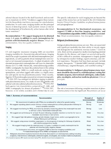
Table 1. Summary of recommendations
Recommendation Strength of Quality of
recommendation evidence
1 We recommend all patients with PPGLs be considered for referral for genetic testing Strong Moderate
2 We suggest repeating plasma and/or 24-hour urinary metanephrines at first postoperative followup to Strong Low
ensure complete resection.
3 We suggest monitoring for PPGL recurrence by annually measuring plasma free metanephrines and/ Strong Low
or 24-hour urinary fractionated metanephrines.
4 We suggest annual followup for at least 10 years following complete resection to monitor for local or Weak Very low
metastatic recurrences or new tumors.
5 We suggest high-risk patients (young, genetic disease, larger tumor, and/or a paraganglioma) be Weak Very low
offered lifelong annual followup.
6 We suggest using annual clonidine suppression or chromogranin A testing for followup of patients Weak Very low
with positive preoperative results.
7 We suggest imaging tests be obtained every 1–2 years, in addition to yearly metanephrines for Weak Very low
patients with biochemically negative disease.
8 For biochemical recurrence, we suggest CT/MRI as first-line imaging modalities, and Weak Very low
123 I-metaiodobenzylguanidine (MIBG) scintigraphy as second-line.
9 Malignant pheochromocytoma treatment should be discussed in a multidisciplinary setting including Clinical principle
surgeons, interventional radiologists, endocrinologists, oncologists, and nuclear medicine physicians.
CT: computed tomography; MRI magnetic resonance imaging; PPGL: pheochromocytomas and paragangliomas.
CUAJ • December 2019 • Volume 13, Issue 12 375