
Page 14 - CANADIAN URINARY DIVERSIONS POSITION STATEMENT
P. 14
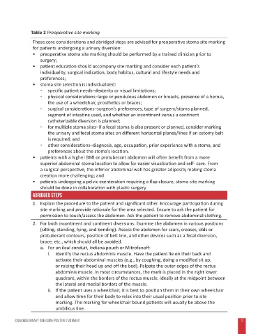
Table 2 Preoperative site marking
                 These core considerations and abridged steps are advised for preoperative stoma site marking
                 for patients undergoing a urinary diversion:
                 •  preoperative stoma site marking should be performed by a trained clinician prior to
                    surgery;
                 •  patient education should accompany site marking and consider each patient’s
                    individuality, surgical indication, body habitus, cultural and lifestyle needs and
                    preferences;
                 •  stoma site selection is individualized:
                       ◦  specific patient needs–dexterity or visual limitations;
                       ◦  physical considerations–large or pendulous abdomen or breasts, presence of a hernia,
                        the use of a wheelchair, prosthetics or braces;
                       ◦  surgical considerations–surgeon’s preferences, type of surgery/stoma planned,
                        segment of intestine used, and whether an incontinent versus a continent
                        catheterizable diversion is planned;
                       ◦  for multiple stoma sites–if a fecal stoma is also present or planned, consider marking
                        the urinary and fecal stoma sites on different horizontal planes/lines if an ostomy belt
                        is required; and
                       ◦  other considerations–diagnosis, age, occupation, prior experience with a stoma, and
                        preferences about the stoma’s location.
                 •  patients with a higher BMI or protuberant abdomen will often benefit from a more
                    superior abdominal stoma location to allow for easier visualization and self- care. From
                    a surgical perspective, the inferior abdominal wall has greater adiposity making stoma
                    creation more challenging; and
                 •  patients undergoing a pelvic exenteration requiring a flap closure, stoma site marking
                    should be done in collaboration with plastic surgery.
                 Abridged steps
                 1.  Explain the procedure to the patient and significant other. Encourage participation during
                    site marking and provide rationale for the area selected. Ensure to ask the patient for
                    permission to touch/assess the abdomen. Ask the patient to remove abdominal clothing.
                 2.  For both incontinent and continent diversions. Examine the abdomen in various positions
                    (sitting, standing, lying, and bending). Assess the abdomen for scars, creases, olds or
                    protuberant contours, position of belt line, and other devices such as a fecal diversion,
                    brace, etc., which should all be avoided.
                    a.  For an ileal conduit, Indiana pouch or Mitrofanoff:
                        i.  Identify the rectus abdominis muscle. Have the patient lie on their back and
                           activate their abdominal muscles (e.g., by coughing, doing a modified sit up,
                           or raising their head up and off the bed). Palpate the outer edges of the rectus
                           abdominis muscle. In most circumstances, the mark is placed in the right lower
                           quadrant, within the borders of the rectus muscle, ideally at the midpoint between
                           the lateral and medial borders of the muscle.
                        ii.  If the patient uses a wheelchair, it is best to position them in their own wheelchair
                           and allow time for their body to relax into their usual position prior to site
                           marking. The marking for wheelchair bound patients will usually be above the
                           umbilicus line.


        CANADIAN URINARY DIVERSIONs POSITION STATEMENT                                                             13 13