
Page 15 - CUAJFeb2023
P. 15
Guideline: Adrenal incidentaloma
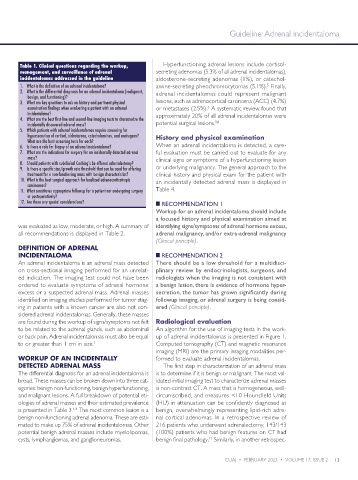
Table 1. Clinical questions regarding the workup, Hyperfunctioning adrenal lesions include cortisol-
management, and surveillance of adrenal secreting adenomas (5.3% of all adrenal incidentalomas),
incidentalomas addressed in the guideline aldosterone-secreting adenomas (1%), or catechol-
5
1. What is the definition of an adrenal incidentaloma? amine-secreting pheochromocytomas (5.1%). Finally,
2. What is the differential diagnosis for an adrenal incidentaloma (malignant, adrenal incidentalomas could represent malignant
benign, and functioning)?
3. What are key questions to ask on history and pertinent physical lesions, such as adrenocortical carcinoma (ACC) (4.7%)
5
examination findings when evaluating a patient with an adrenal or metastases (2.5%). A systematic review found that
incidentaloma? approximately 20% of all adrenal incidentalomas were
4. What are the best first-line and second-line imaging tests to characterize the
3,6
incidentally discovered adrenal mass? potential surgical lesions.
5. Which patients with adrenal incidentalomas require screening for
hypersecretion of cortisol, aldosterone, catecholamines, and androgens? History and physical examination
What are the best screening tests for each?
6. Is there a role for biopsy of an adrenal incidentaloma? When an adrenal incidentaloma is detected, a care-
7. What are the indications for surgery for an incidentally detected adrenal ful evaluation must be carried out to evaluate for any
mass? clinical signs or symptoms of a hyperfunctioning lesion
8. Should patients with subclinical Cushing’s be offered adrenalectomy?
9. Is there a specific size/growth rate threshold that can be used for offering or underlying malignancy. The general approach to the
treatment for a non-functioning mass with benign characteristics? clinical history and physical exam for the patient with
10. What is the best surgical approach for localized adrenocorticotropic an incidentally detected adrenal mass is displayed in
carcinomas?
11. What constitutes appropriate followup for a patient not undergoing surgery Table 4.
or postoperatively?
12. Are there any special considerations? █ RECOMMENDATION 1
Workup for an adrenal incidentaloma should include
a focused history and physical examination aimed at
was evaluated as low, moderate, or high. A summary of identifying signs/symptoms of adrenal hormone excess,
all recommendations is displayed in Table 2. adrenal malignancy, and/or extra-adrenal malignancy
(Clinical principle).
DEFINITION OF ADRENAL
INCIDENTALOMA █ RECOMMENDATION 2
An adrenal incidentaloma is an adrenal mass detected There should be a low threshold for a multidisci-
on cross-sectional imaging performed for an unrelat- plinary review by endocrinologists, surgeons, and
ed indication. The imaging test could not have been radiologists when the imaging is not consistent with
ordered to evaluate symptoms of adrenal hormone a benign lesion, there is evidence of hormone hyper-
excess or a suspected adrenal mass. Adrenal masses secretion, the tumor has grown significantly during
identified on imaging studies performed for tumor stag- followup imaging, or adrenal surgery is being consid-
ing in patients with a known cancer are also not con- ered (Clinical principle).
sidered adrenal incidentalomas. Generally, these masses
are found during the workup of signs/symptoms not felt Radiological evaluation
to be related to the adrenal glands, such as abdominal An algorithm for the use of imaging tests in the work-
or back pain. Adrenal incidentalomas must also be equal up of adrenal incidentalomas is presented in Figure 1.
to or greater than 1 cm in size. 1 Computed tomography (CT) and magnetic resonance
imaging (MRI) are the primary imaging modalities per-
WORKUP OF AN INCIDENTALLY formed to evaluate adrenal incidentalomas.
DETECTED ADRENAL MASS The first step in characterization of an adrenal mass
The differential diagnosis for an adrenal incidentaloma is is to determine if it is benign or malignant. The most val-
broad. These masses can be broken down into three cat- idated initial imaging test to characterize adrenal masses
egories: benign non-functioning, benign hyperfunctioning, is non-contrast CT. A mass that is homogeneous, well-
and malignant lesions. A full breakdown of potential eti- circumscribed, and measures <10 Hounsfield Units
ologies of adrenal masses and their estimated prevalence (HU) in attenuation can be confidently diagnosed as
is presented in Table 3. The most common lesion is a benign, overwhelmingly representing lipid-rich adre-
1-4
benign non-functioning adrenal adenoma. These are esti- nal cortical adenomas. In a retrospective review of
mated to make up 75% of adrenal incidentalomas. Other 216 patients who underwent adrenalectomy, 143/143
potential benign adrenal masses include myelolipomas, (100%) patients who had benign features on CT had
cysts, lymphangiomas, and ganglioneuromas. benign final pathology. Similarly, in another retrospec-
11
CUAJ • FEBRUARY 2023 • VOLUME 17, ISSUE 2 13